Basic Information & Functions
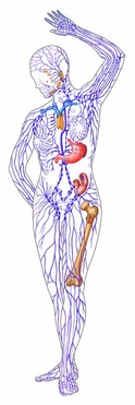
The lymphatic system spans the entire body and serves as a sort of drainage system that fights infections and cancers whiel carrying nutrients to cells. It can be described (vaguely) as the system that is as a network of tissues, ducts and organs that plays a vital role in immunity (an individual's ability to resist and/or overcome the effects of a harmful agent) via a partnership with the circulatory system. The lymphatic system itself is comporable to the circulatory system in that is like a tree with braches called lymphatic vessels that cary the clear fluid lymph to the lymph nodes from tissues all over the body (everything in the body is bathed in lymph). There are about 100 lymph nodes placed strategically inside the body (they are about the size and shape of a jelly bean). Lymph carries harmful substamce from organs and other tissues to the lymph nodes. The lymph is forced to percolate, or pass through, the connective honeycomb tissue of the lymph node. This creates a filter. The bad substance in the lymph are destroyed there. The lymphatic system, specifically lymph nodes, create specialized proteins called antibodies that respond to foreign invaders brought to them by the lymph. Responses can either be specialized, tailored to destoy certain pathogens, or general, protect the body from inavders in general. The making of antibodies begins when the lymph brings a foreign invader to the nodes. This stimulates the formation of white blopod cells called lymphocytes (B cells). These antibodies combine with the antigen in order to neutralize it. This process serves as an explanation as to why your lymph nodes swell when you are fightining an infection; they become enlarged because they have to producing antibodies.
Breaking it down: Level 1: Involved Organs
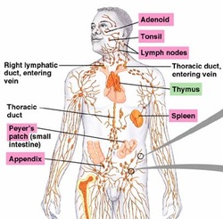
Tonsils are clumps of lymphoid tissue located on either side of the throat that are embedded in the roof of the mouth. Adenoids are a single clump of the lymphoid tissue in the back of the nose (nasopharynx) in children. In adults they are located in the back of the throat (pharynx) about one inch above the uvula. The tissue that makes these things up is composed of lymphocytes that produce antibodies. They were designed to protect againts the invasion of antigens from the air.
Peyer's patches are lymphoid nodules taht are similar to lymph nodes that are found in the lower part of the small intestine (the ileum of the epithelium). They destroy bacteria that is trying to breach the wall of the small intestine and enter the bloodstream. They also generate memory lymphocytes for long term immunity. They are a part of mucosa-associated lymphoid tissue or MALT tissue group.
The thymus gland is an organ that lies beneath the breast bone. It process a type of lymphocyte called T-lymphocyte (T-cells) that aid in the recognition and destruction of bacteria, viruses and abnormal cell growth (cancer). It also produces the horomone thymosin which promotes the growth of lymphocytes and lymphoid tissue in the body.
The spleen, located in the upper left part of the abdomen below the diaphragm, is a filter that creates lymphocytes for the destruction and recycling of red blood cells. It also serves as a blood reservoir that supplies blood to the body when it is suddenly need (i.e. a cut). It is also where white blood cells capture and destroy antigens.
The actual function of the appendix is unknown. However, it is composed of lymph tissues. It is loacted at the end of the cecum, a part of the large intestine.
Bone marrow, though not an organ, is a vital part of the lymphatic system because it is where blood cells are produced. They play an importan role in immunity and are involved with the lymphatic system.
Breaking it down: Level 2: Lymph nodes
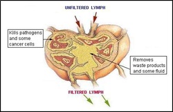
Lymph nodes come in many variations of the jelly bean size mentioned above but a standard nod eis about one inch long and the size of a bean. When a human is born, they have about 600-700 lymph nodes. As we age, the number of lymph nodes decreases to about 100. They are covered in a capsule and are divided into the cortex and medulla. The cortex holds the lymphocytes known as B-cells which produce antibodies that circulate. The rest of the cortex contains T-cells that circulate the lymphatic system searching and destroying pathogens. The medulla is where a macrophage, another type of leukocyte, attach the the fibers of the node. Lymph node are what filter lymph.
Breaking it down: Level 3: Lymph
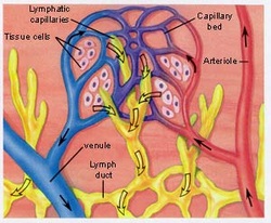
Lymph is a virtually colorless fluid similar to blood plasma in composition that carries harmful substances from tissues to lymph nodes to be filtered.This is possible due to pores in capillaries that allow the lymph to escape the circulatory system. After it has been filtered, the lymph is then reintergrated with the blood through veins in the neck. It is now more likely to be altered in composition because it has picked up lymphocytes and other proteins. Lymph, before it is porcessed by a node, is an interstitial fluid which means it serves as a vessel for nutrients and oxygen to reach and be disposed of by cells. Substances are constantly being interchanged between lymph and cells.
Second helping: Lymphadema
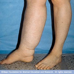
Lymphadema is a rare, heritable and incurable disease that causes swelling (most often in the arms and legs, usually only on one side) caused by problems with the development of lymph vessels. It occurs primarily in women. The swelling cahracteristic of lymphadema is the result of a blockage in the lymph vessels. The blockage impedes the drainage of lymph and the fluid begins to pool and cause enlargement of the affected area. This swelling can range from barely noticable to extreme swelling that restricts motion. The affected limb feels heavy and a general sense of discomfort is felt. It can either be primary, meaning it occurs on its own, or secondary, meaning it occurs because of another disease. Primary causes include Milroy's disease (congenital lymphadema), Meige's disease (lymphadema praecox), and late-onset lymphadema (lymphadema tarda). Secondary lymphadema is often cause by surgery, radiation treatment, cancer and infection. Severe cases left untreated can result in a rare type of soft flesh cancer called lymphangiosarcoma. Infection of affected limbs is common. Treatment includes certain exercise that require movement of the affected limb, wrapping the arm or leg, massage, pneumatic compression and compressive garments. It is important to note that this disease can be psychologically distressing due to the visibility of the swelling of the limb and having to cope with the lack of a cure.
Image Citations
http://www.dorlingkindersley-uk.co.uk/nf/ClipArt/Image/0,,239033_1582321_,00.html
http://faculty.irsc.edu/FACULTY/TFischer/bio%202%20files/Bio%202%20resources.htm
http://www.hls-herbs.com/health_condition/Lymph-Drainage.html
http://www.biosbcc.net/doohan/sample/htm/COandMAPhtm.htm
http://www.mayoclinic.com/health/medical/IM03018
http://faculty.irsc.edu/FACULTY/TFischer/bio%202%20files/Bio%202%20resources.htm
http://www.hls-herbs.com/health_condition/Lymph-Drainage.html
http://www.biosbcc.net/doohan/sample/htm/COandMAPhtm.htm
http://www.mayoclinic.com/health/medical/IM03018
